1. A Physical Barricade in Pancreatic Cancer
A couple of weeks ago, I met Kenneth Olive at Columbia, who spoke about the following story coming out of his lab:

Basically, Olive looked at the (dismal) fact that pancreatic cancer seems to be resistant to almost every kind of chemotherapy. The current standard of care, gemcitabine, extends survival by about two weeks. He started studying how pancreatic cancer in mice. As expected, treating mice with pancreatic tumors with gemcitabine didn't do much. However, Olive noticed that if he took pancreatic cancer cells out, grew them in a dish, and transplanted them into the skin of another mouse, they suddenly became sensitive to gemcitabine. So he wondered if maybe the issue wasn't that the cancer cells were resistant to the drug, but rather that the drug just couldn't get into the tumor.
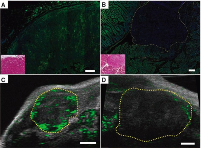
So he looked at tumors that were either in the pancreas (b/d, left) or transplanted subcutaneously (a,c). He found that doxorubicin (used because it autofluoresces green) rapidly entered transplanted but not native tumors. When he imaged the tumors by ultrasound (c,d), he found similarly that blood flow into native tumors was minimal.
 It turns out that pancreatic adenocarcinoma is characterized by a very potent proliferation of fibroblasts. Olive hypothesized that the stromal cells might actually create a physical barrier against drug entry. So he treated mice with a small molecule inhibitor of smoothened, a member of the sonic hedgehog pathway that is critical for fibroblast proliferation. And indeed, he found that when fibroblast growth was inhibited, the pancreatic tumors became more vascularized, more perfused with chemotherapeutic drugs, and the mice lived longer. This drug is now in accelerated phase II trials. It's not a cure, but it is a great example of out-of-the-box thinking.
2. Looking deeper into B cell development
I don't really know that much about B cells, but I know enough to appreciate my pal Gabriel Victora's landmark study in Cell last month, titled, "Germinal center dynamics revealed by multiphoton microscopy with a photoactivatable fluorescent reporter." Basically, B cells undergo antigen recognition in lymph nodes where they assemble into histologically visible cellularly dense structures called 'germinal centers.' Germinal centers are composed of a 'light zone' and 'dark zone' based on how they look on histology slides. When B cells see antigen, they begin to undergo a process known as 'affinity maturation' in which they undergo rapid mutation (somatic hypermutation) of the antigen-recognizing sequences on their immunoglobulin receptors. Somehow, this results in survival of only high affinity B cell clones in a process that somehow involves both T cells and follicular dendritic cells. How this relates to creation and maintenance of the light and dark zones of the germinal center was suspected, but not known, until this study from Victora and colleagues.
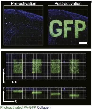
First and foremost, the authors developed a technique in which they could use two-photon microscopy to sensitively apply a light stimulus to specific regions of the lymph node. That light stimulus would then uncage a photoactivatable fluorescent reporter, GFP, only in cells receiving the stimulus (specificity shown awesomely to the right). Read here to see why two-photon microscopy is critical to this process - basically, it can more efficiently target light to a single plane.
Victora and colleagues used this technique to differentially activate only cells in the light or dark zone of germinal centers. Keep in mind - this is the first time that light and dark zone cells could be discriminated while still alive. Using this technology, they were able to assess differential surface protein expression as well as gene expression in light and dark zone B cells.

What they found was that the dark zone B cells expressed more genes related to mitosis (consistent with the idea that cells in the dark zone are rapidly proliferating) whereas light zone B cells expressed more molecules related to activation and apotosis, suggesting that this is where B cells undergo antigen recognition and selection.
Next, Victora and colleagues used this photoactivatable technique to track migration of B cells between the dark and light zone.

They found that B cells moved rapidly from the dark zone to the light zone, repopulating the light zone within about six hours, but more slowly from the light zone to the dark zone. Finally, they showed that survival of B cells in the light zone was critically dependent on presentation of antigen on MHC-II molecules to T cells in the light zone. Based on this, Victora and colleagues presented an integrated model for affinity maturation of B cells in germinal centers. B cells acquire antigen by moving from the dark zone to the light zone where they migrate along follicular dendritic cells. As they divide and their Ig receptors undergo SHM, the cells that develop higher affinity receptors are able to capture more antigen, present more antigen to T cells, and therefore receive more T cell help. These are the B cells which survive, return to the dark zone, and undergo proliferation. Kudos to Gabriel and the members of the Dustin and Nussenzweig labs who worked on this super-cool project.
No comments:
Post a Comment